


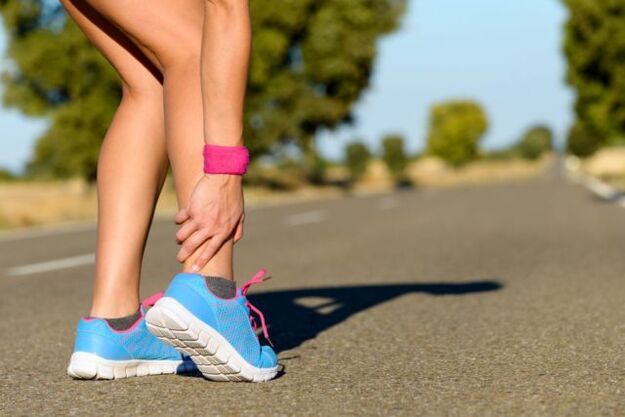
The ankle joint is often injured due to heavy loads. Such a diagnosis of ankle joint disease is not uncommon. It is set regardless of the patient's age and sex. What is ankle joint disease and how is it treated?
What is it?
There is a terrible load on the ankle. Its function is to keep the body upright. Thanks to him, a person walks and runs. With a violation of the ankle system, it is difficult to lead a familiar lifestyle. What interrupts the work of the ankle?
Ankle joint, what is it? This is a chronic joint disease, which is characterized by a degenerative process. In the cartilage of the joints, irreversible processes are activated, which leads to formidable complications.
The ankle joint develops gradually. Healthy joint surfaces are elastic and smooth. They provide cushioning under heavy loads and glide smoothly while driving. With pathology, tissue nutrition and metabolism are disturbed. The surface of the joint becomes less elastic and rough. During exercise, the flesh fibers come into contact with each other, leading to inflammation. When lifting weights, the main load falls on the bones, which threatens degenerative disorders.
Lack of treatment leads to more serious disorders. At stage 3-4, cartilage and tissue damage is observed. The bursa is inflamed. The joint becomes unstable. Support function is violated. All these violations in total lead to the fact that movement becomes impossible.
Arthritis (degenerative joint disease) is one of the most common joint diseases, affecting quite a few people.
Causes and risk factors
What is ankylosing spondylitis, we have classified it together. Now let's find out what its root cause is. Ankle joint is considered a disease of old age. This is due to age-related changes in the body. Cartilage becomes thinner, bones become unstable and brittle. However, in the past decade, the diagnosis of ankle joint has become much younger. Such statistics are disappointing, as many patients ignore the first signs of the disease. Late diagnosis always threatens the development of serious complications.
Trigger factors include:
- dislocation;
- bruises;
- inflammatory diseases;
- injury;
- excess weight;
- metabolic impairment;
- intolerable physical activity;
- wearing uncomfortable shoes;
- autoimmune and endocrine diseases;
- bone necrosis.
Clinical symptoms
The ankle joint is identified by the following features:
- Pain. The disease is initially mild and occurs after walking or exertion. Sometimes a person is in an uncomfortable position. With the progression of the pathological process, the pain syndrome intensifies and the anxiety rests.
- Swelling and inflammation. These signs appear against the background of trauma and dislocation. Body temperature in the affected area increases.
- Click. When the ankle is affected, the puncture is "dry" and causes pain.
- Dislocation or misalignment. Due to thinning of cartilage tissue and degeneration, joints become unstable. Bones can shift and fall out of the joint capsule. These changes cause acute attacks of pain.
- Stiffness. When cartilage tissue is replaced, the joint no longer functions properly, negatively affecting its mobility.
- Joint deformity. This symptom appears in 3-4 stages of joint disease. Osteoarthritis also leads to bent ankles.
If any of these symptoms appear, see a doctor immediately. Initiating treatment promptly is a step towards recovery.
Arthritis of the feet and ankles is characterized by a slow progression, with clinical manifestations developing gradually over several years.
Classification and stages
The disease develops in different ways. In some patients, several years pass from the first signs to the final stage, in others a rapid development of the disease is observed. The speed depends on the age and health of the patient, when the treatment is started. The symptoms of dry ankle joint become brighter as the disease progresses.
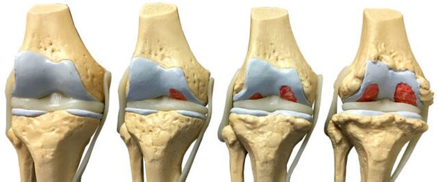
There are four stages of joint disease:
- The early stages often go unnoticed. Morning stiffness and ankle pain are sometimes present after intense exertion. When the feet move, a characteristic crunching sound is heard. Pathological changes are not yet visible on x-rays, but cartilage destruction has already begun.
- Morning stiffness becomes persistent. It takes 20-30 minutes to develop a leg. Sometimes a limp occurs. Grade 2 ankle osteoarthritis is noted on the graphic image by the growth of bone tissue, the displacement of the bone.
- The symptoms in the 3 stages are all obvious. Worry about pain is not only after a load but also when resting. It is difficult for the patient to perform without pain medication. The lameness increased. Crutches may be needed. The affected joint is swollen and deformed. The ankle muscles are atrophied. X-ray showed that the joint space was narrowed, the formation of bone sockets, compression sockets.
- Stage 4 is the hardest. It develops due to lack of treatment. Cartilage is destroyed, the surfaces of joints are fused. Can't walk anymore.
With the development of ankle osteoarthritis, there is a gradual change in the cartilage and bone tissue of the joint surface.
Diagnose
Diagnosis of ankle joint disease is based on clinical symptoms and information obtained on examination. Laboratory studies are considered ineffective, since there are no special tests that can detect pathology. During remission, all parameters are within the normal range, with an exacerbation of the disease, clinical blood tests will show high levels of C-reactive protein and ESR. These indicators indicate that the pathological process has begun.
To confirm the diagnosis, instrumental methods are used:
- X-ray machine;
- Magnetic resonance imaging;
- Supersonic;
- Bone scan;
- diagnosis of joint perforation.
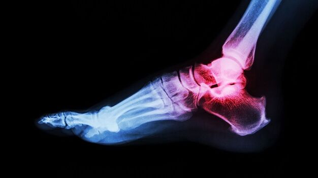
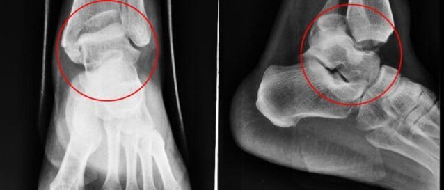
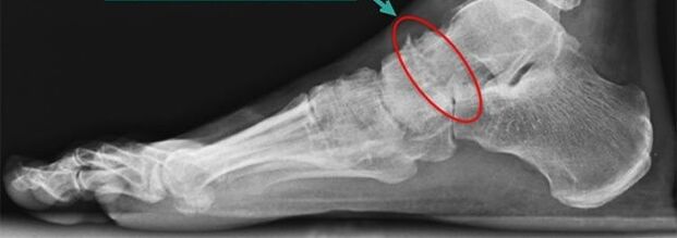
Simple X-ray
Plain radiography is the most reliable and effective method for the diagnosis of diseases occurring in the musculoskeletal system. The principle of manipulation is the different absorption of X-rays by muscle tissues. Soft tissues allow X-rays to pass through, but hard tissues absorb them. X-rays allow you to diagnose both the disease itself and its consequences.
A conventional X-ray is an examination in which a small amount of X-rays are passed through the person's body or part of the body.
Snapshots allow you to see:
- Condition of the bony surface in the joint.
- The shape, size and arrangement of the structures in the joint are relative to each other.
- Condition of fabric.
- The size of the joint space.
These indicators help the doctor determine the type and extent of joint damage. If the data are not enough, then the doctor will appoint other studies.
With ankle arthritis, X-rays are taken in three projections:
- beside;
- backside;
- back with one foot moving inward.
The disease is characterized by the following changes:
- reduce the common space;
- the presence of bone-forming substances;
- bone cartilage replacement (subchondral sclerosis);
- small void in the peristaltic part.
Benefit from nuclear
Nuclear magnetic resonance (NMR) as a diagnostic method allows you to study parts of the body where water is present. The image shows bones as dark, because they contain less water, but muscle tissue, discs, and nerves are shown lighter. MRI allows you to detect the smallest changes in the structure of bone and joint tissue. The study is also indicated for patients before joint restoration. YMG has one drawback - high price.
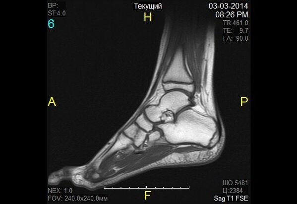
At nuclear magnetic resonance, a change in the properties of the hydrogen molecule is observed under the influence of a strong magnetic field.
Magnetic resonance imaging
Magnetic resonance imaging (MRI) is an alternative diagnostic method that allows you to carefully examine the ligamentous structure of joints, muscles, and cartilage tissue. With the help of an MRI, the doctor will evaluate the condition of the lower leg joints. Based on the survey data, the pathology is revealed at an early stage of development.
The diagnostic principle is based on exposure to radio waves and strong magnetic radiation. The magnetic field used is not hazardous and poses no health hazard.
MRI is contraindicated in case of mental disorders, during pregnancy and when metal objects are present in the human body.
When diagnosing arthropathy of the ankle, classical (closed-type) MRI machines are used, as they have better image quality. An MRI machine is a large cylindrical tube, surrounded by magnets. The patient lies down on a special table. The ankle is fixed with a special coil. The procedure takes about 30-40 minutes. This study is completely painless. The patient may feel heat in the lower leg area.
Supersonic
Ultrasound examination has been widely used in medicine since the 90s of the twentieth century. This technique has proven to be good at making accurate diagnoses. An ultrasound is also done to look for ankle arthritis.
Today, ultrasound examination is of no particular importance in the diagnosis of osteoarthritis, as it does not allow for a complete study of damaged joints.
The device on which the research was carried out generates waves at extremely high frequencies. The waves are reflected from the tissues and recorded on a monitor. Based on the resulting image, the doctor determines the type of pathology. To make the image on the screen clear, a special gel is used. It eliminates air gaps and helps the sensor glide better.
The ultrasound examination does not harm the patient, so the procedure can be repeated many times. Advantages of ultrasound also include low cost and high accuracy.
The following indicators are a clear sign of joint disease:
- thinning cartilage;
- the presence of bone growth;
- accumulation of effusion in the joint cavity (bursitis);
- loss of cartilage space.
Bone scan
Scintigraphy is a highly accurate study, using isotopes, that can detect pathological changes in bone. Doctors divide the foci of disease into "cold" and "hot". In the first case, we are talking about regions in which there are no isotopes. These areas are poorly supplied with blood and they are not visible during the scan. The "cold" area is where the malignancy is affected. In "hot" regions, isotopes quickly accumulate, and they look very bright when scanned. Such areas indicate the presence of inflammatory processes.
The role of scintigraphy in joint disease is substantial. Research helps to distinguish arthropathy from a number of other diseases when the clinical symptoms are extremely similar.
During a bone scan, a special preparation containing specially labeled atoms is injected into the body.
Based on the results of the scan, the doctor gives a clinical prognosis and determines the treatment regimen. The only limitation of the study is the high cost. Scintigraphy is performed using special equipment, and unfortunately, not all medical facilities can afford it.
Although a radionuclide scan is a safe procedure, it still has some contraindications:
- pregnancy;
- lactation period;
- use barium-containing drugs.
When radioactive material is injected, some patients experience allergic reactions in the form of itching and rashes. These side effects are not dangerous and go away on their own in a short time.
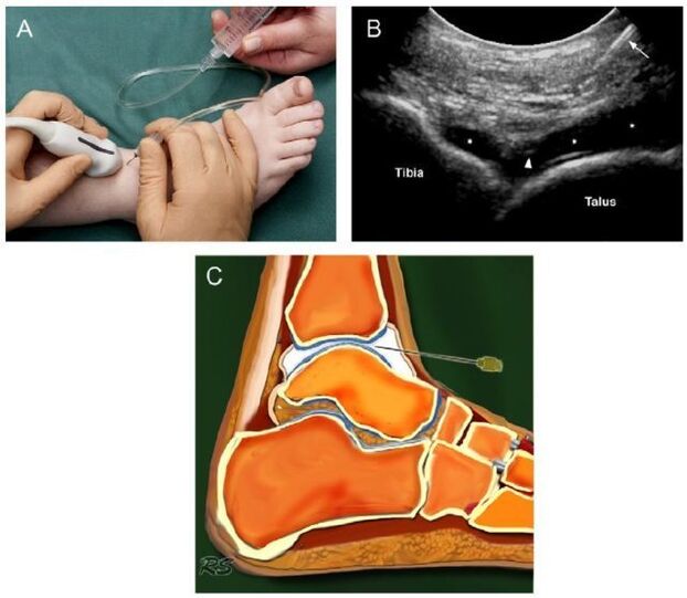
Joint perforation
Arthroscopy is a diagnostic procedure in which a needle is inserted into the joint cavity to collect joint fluid. This liquid is then sent for further study. Based on the data obtained, the doctor makes a conclusion about the nature of the disease and the stage of its development.
At first glance, puncture is a simple trick, but it is not. The removal of fluid from the joint capsule requires exceptional precision in the doctor's movements. The bursa is very thin and an awkward movement will hurt it. As a result, an inflammatory process develops. Potential risks also include infection. It is not difficult to introduce infection into the joint capsule through poorly sterilized instruments.
Manipulation techniques are different for each joint. When collecting joint exudate from the ankle, a puncture is made anteriorly, between the outer ankle and the tendon of the elongated nerve.
Intraarticular fluid sampling allows laboratory analysis and excludes inflammatory arthritis.
Basic principles of treatment
Once the diagnosis of ankle arthritis is confirmed, the symptoms should not be long in coming. Treatment is started immediately. Further prognosis depends on a well-chosen treatment regimen and timing of initiation.
Arthritis is an insidious disease. It cannot be completely cured. The goal of therapy is to stop degenerative processes and prolong remission. For this purpose, the doctor prescribes drugs, physiotherapy, massage, therapeutic gymnastics and folk remedies. If the conditions are met, it can be counted as positive, otherwise the disease progresses.
Drug treatment for joint disease
Depending on the therapeutic effect, the drug is divided into several groups:
- Anti-inflammatory or pain relievers. This group of drugs is intended to eliminate the focus of inflammation and relieve pain. The earlier anti-inflammatory treatment is started, the greater the chance of saving the joint. Medicines in this group can be produced in the form of tablets and ointments.
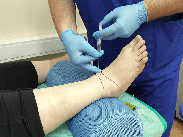
- Glucocorticoids. These drugs are prescribed when the above funds are not effective. They are produced in the form of a solution for injection. The drug is injected directly into the joint.
- Chondroprotectors. Designed to slow cartilage destruction.
The treatment regimen and dosage are selected by the doctor, based on the severity of symptoms, the patient's age, the presence of concomitant diseases and other factors. Self-medication is very dangerous and often aggravates the situation, since many drugs have a number of side effects and contraindications of their own.

Features of radical treatment
If conservative treatment is not successful, doctors are forced to resort to radical treatment (surgical intervention). This activity is also displayed when:
- secondary (post-traumatic) and primary joint grade 3-4;
- matching complications;
- constant and severe pain in the ankle, radiating down to the knee;
- severe limp;
- paralysis and paralysis of the leg muscles;
- violation of the flexor-extensor function of the joint;
- violation of the ability to support the foot.
Surgical intervention is contraindicated if:
- patients under 12 years old;
- fistula is found in the joint;
- patients with a history of diabetes, heart failure;
- Infectious diseases have been found in the proposed intervention area.
Traditional treatment
Doctors believe that the treatment of joint diseases should be done completely under the supervision of a specialist, but they do not deny the positive effects of folk remedies. Alternative medicine acts as an effective prophylactic that eliminates symptoms and maintains remission.

Folk remedies are symptomatic treatment for foot joint disease.
It is advisable to coordinate home treatment with your doctor to avoid side effects and complications.
Traditional healers recommend treating ankle sprains with:
- Burdock. Wash the burdock leaves with soap and water. Apply the leaves with the soft side to your skin. Secure the top with tape or cling film. It is better to keep the gauze all night.
- Sea salt. Chop the salt in the pan. Pour it into a linen bag and attach it to your ankle. Hold the bag until the salt has cooled. Heat relieves pain. Sand, lentils, buckwheat are also used instead of salt.
- Lilacs. Pour perfume over the lilac flowers. Leave tincture in a dark and cool place for 10-14 days. Rub the affected area morning and evening.
- Eggshell. Grind the pods in a coffee grinder. Take the formed dough for ½ tsp. before eating.
Do not forget that treatment with folk remedies should not be the only remedy. Complex treatment includes medication, therapeutic exercise, massage, physiotherapy, spa treatment. In severe cases, the doctor uses radical measures - surgical intervention.
Surgery
For arthropathy, the following types of manipulation are used in medicine:
- arthrodesis of the joints;
- arthroscopy;
- endogenous drugs.
Arthrodesis is a surgery to immobilize a joint. It is done to return the cost of lost support capacity. The only drawback of surgery is that the bones (tibia and fibula) grow together, resulting in immobility. Arthrodesis is rarely used in medical practice.
Arthroscopy is a minimally invasive procedure. During surgery, the doctor will make small incisions in the joint area and through it, insert an arthroscope (a special tube at the end with a camera attached). With its help, the surgeon carefully examines and evaluates the condition of the structures in the joint. If necessary, damaged joint fragments or blood clots are removed from the joint fluid. This maneuver is less traumatic. The only drawback of arthroscopy is the high risk of recurrence.
Endoscopy is the last resort. It is done with advanced joint disease. Arthroscopy allows you to replace the affected joint partially or completely. As a prosthetic product, innovative prostheses with modern mechanics are used. An artificial joint has a lifespan of 10 to 20 years.
Power feature
To achieve a favorable result, drug therapy is supplemented with dietary therapy. Nutritionists have developed a special diet to avoid exacerbation of the disease, while providing a full range of vitamins and nutrients needed by the body. Diet for overweight patients plays a special role. Since obesity is one of the causes leading to the development of joint disease, weight management is an integral part of treatment.

Patients need to review some of their habits in daily life, these habits contribute to and stimulate the progression of foot joint disease.
Nutritionists recommend compliance with the following nutritional conditions:
- Eat often and in small portions.
- Drink at least 2 liters of fluid per day.
- Give up sweets and salt.
- Last meal no later than 18. 00.
- Dishes are allowed to be steamed, boiled or grilled.
The main task of the diet for joint disease is balanced and fortified nutrition. Fasting is out of question. Extreme diets and detoxes do more harm than good. Calcium is eliminated from the body, necessary for the repair of cartilage. A dietitian will help you compose a daily diet.
With arthrosis, it is allowed to eat cereals, pasta, dairy products, cheeses, legumes, vegetables, fruits, rye bread, dried fruit, nuts, fish, poultryhold. Heavy and fatty side dishes, foods containing dyes and flavorings, as well as pickles, sauces, smoked meats, fatty broths, pies, condiments, sauces, chocolate, ice cream, coffeeand alcohol are prohibited.
Prevention of joint disease
To avoid the development of ankle arthritis, doctors recommend taking preventive measures:
- wear comfortable shoes without heels;
- adhere to a diet and drink enough fluids;
- seasonal vitamin and mineral supplements;
- swimming;
- walk more in the fresh air;
- eliminate excessive stress on the legs;
- avoid hypothermia;
- be examined by a doctor in a timely manner.
With existing joint disease, you should make lifestyle adjustments:
- To reject bad habits. It has been shown that they cause stagnation of blood in tissues and accelerate the destruction of cartilage.
- Do a set of exercises to warm up your ankles.
Forecast
Arthritis is a progressive disease. If left untreated, it can lead to irreversible consequences and complete immobilization of the joint. Early diagnosis of pathology allows you to do without radical measures. The drug is able to suspend the pathological process and alleviate the patient's condition. The fight against this disease in the early stages is uncomplicated.